
Pediatric chest X-ray AI that shows its work.
Two explainable tools, both trained on children's imaging: one for thoracic findings and one for support-device placement.

An explainable second opinion for pediatric chest findings
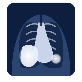
PedXR Pathology flags abnormal findings on pediatric chest X-rays and highlights the image regions that led to each one, so clinicians can check the evidence before using it in their decision.
Findings on the roadmap
What makes it different
Landmark-anchored findings
Each output is tied to named anatomy, such as the carina, diaphragm or clavicles, so the reader can see the reference point and not just the result.
Pediatric-native training
Trained on children's imaging, unlike the 97%+ of FDA radiology AI that used no pediatric data. We validate performance by age group.
Built for small datasets
Pediatric data is scarce. Our architecture is designed to learn from limited pediatric datasets and stay interpretable.
Precision medicine mode
Adapts to each hospital's imaging hardware and protocols, which reduces the site bias that generic AI introduces.
Catch misplaced tubes and lines before they cause harm
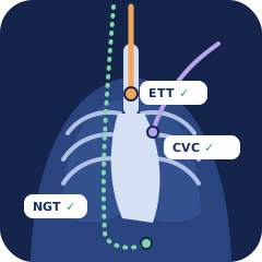
PedXR Tool locates critical support devices on pediatric X-rays and checks their placement against anatomical landmarks, with a visual explanation for every check.
Devices detected & verified
What the AI checks
Endotracheal tube
Confirms midline placement, with the tip below the clavicles and above the carina, where the trachea splits into the bronchi.
NG / OG tubes
Checks that the tube ends below the diaphragm in the stomach before feeding begins.
Central venous catheters
Tracks access through the internal jugular or subclavian vein and confirms the tip sits at the cavoatrial junction.
Umbilical catheters (UAC / UVC)
Critical in neonatal care. Checks UAC and UVC tips against key vascular landmarks to help prevent injury in newborns.
Clinicians told us: no black boxes.
In 20+ NSF I-Corps interviews across 7 organizations, the same concern kept coming up: radiologists won't adopt AI they can't understand. So PedXR is designed to reason the way a pediatric radiologist does.
See the evidence
Highlighted regions show exactly which part of the image drove each finding.
Follow the logic
Findings reference real anatomy, matching how specialists explain a read.
Make the call
The clinician reviews the evidence independently. PedXR supports the decision but doesn't make it.
Plugs into PACS. No new hardware.
Works where you read
Designed to integrate through PACS vendor marketplaces using standard DICOM.
Secure cloud
HIPAA-compliant, auto-scaling infrastructure that can handle winter surges.
Built for FDA review
Built under a design-controlled quality system, targeting a Class II 510(k) pathway.
Interested in a clinical pilot?
We're partnering with select pediatric and community health systems for early validation of the PedXR Suite.